|
|
Molecule of the Month December 2004 |
|
| The PEP-Binding Domain of Enzyme I from Thermoanaerobacter tengcongensis | ||
|
||
|
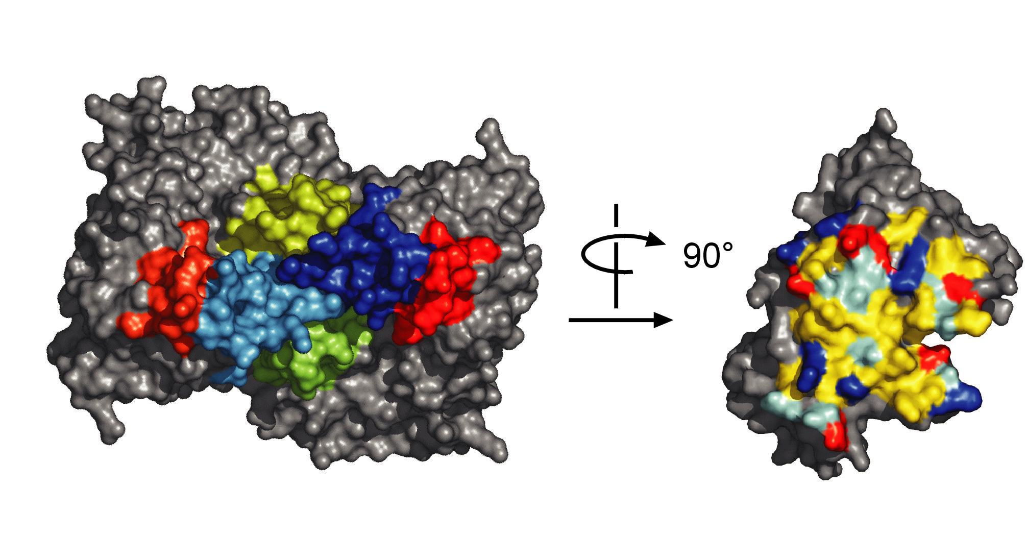
Space filling representation of the dimer and intersubunit contact area of one monomer. The contact area measures 3'750 Å2. It is exceptionally large for a dimerization surface which usually are of the order of 1200-2400 Å2. Three large extensions of the TIM-barrel fold are colored in red, blue and green, apolar, polar, basic and acidic residues in yellow, cyan, blue and red. The protein was purified and crystallized by Philipp Schneider and the structure was solved by Anselm Oberholzer and Mario Bumann from the group of Prof. Bernhard Erni. References:
|
||